“We have not wings we cannot soar; but, we have feet to scale and climb, by slow degrees, more and more the cloudy summits of our time”
lower limb exercises
LOWER LIMB EXERCISES
Hamstrings
3 muscles make up the hamstring muscle group-the bicep femoris, semitendinosus and semimembranosus.

Location
Actions
Injuries
Quadriceps
The quadriceps is a large muscle group that includes the four prevailing muscles on the front of the thigh.
It is subdivided into four separate portions or ‘heads’, which have received distinctive names: Rectus femoris occupies the middle of the thigh, covering most of the other three quadriceps muscles, the Vastus Lateralis, Vastus Medialis and Vastus Intermedius.

Location
Actions
Calf muscles
The calf muscles are made up of one large muscle the gastrocnemius, and a smaller muscle, the soleus, which lies under the gastrocnemius.

Location
Actions
Injuries
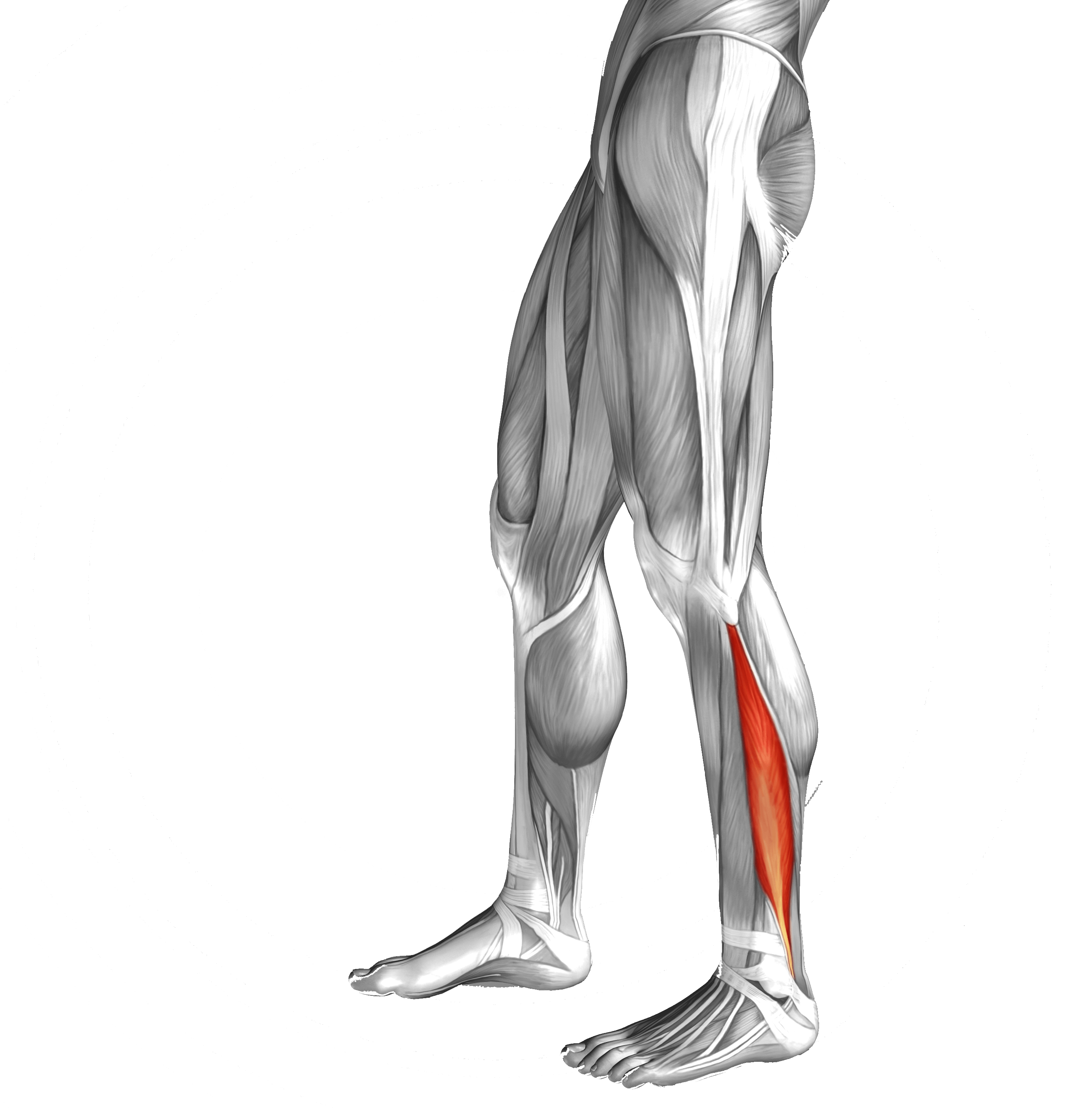
Tibialis anterior

Location
Actions
Injuries
Whenever the tibialis anterior muscle contracts or is stretched, tension is placed through the tibialis anterior tendon. If this tension is excessive due to too much repetition or high force, damage to the tendon can occur. Tibialis anterior tendonitis is a condition whereby there is damage to the tibialis anterior tendon with subsequent inflammation and degeneration.
Patients with tibialis anterior tendonitis usually experience pain at the front of the shin, ankle or foot during activities which place large amounts of stress on the tibialis anterior tendon These activities may include walking or running excessively (especially up or down hills or on hard or uneven surfaces), kicking an object with toes pointed (e.g. a football), wearing excessively tight shoes or kneeling.
Ankle
The ankle joint consists of three bones: the tibia, the fibula, and the talus. The ankle ( talocrural joint), is a synovial hinge joint.
Three separate ligaments stabilize the lateral aspect of the ankle joint: the anterior talofibular, calcaneofibular and posterior talofibular ligaments. Medially, support comes from a collective group of ligaments known as the deltoid ligament.

Major Motions of the Ankle Joint
Inversion/Eversion.
Plantarflexion/Dorsiflexion.
Pronation/Supination. In simple terms, pronation occurs when the plantar side of the foot moves toward the floor surface in weight bearing, and supination occurs when the plantar side moves away from the floor surface. Pronation involves abduction, eversion and some dorsiflexion, whereas supination involves adduction, inversion and plantar flexion.
Injuries
Peroneals
The Peroneal muscles are a group of 3 muscles that originate from fibula (lower leg bone) and for this reason, these are also known as fibularis muscles.

Location
Actions
Both peroneal longus and brevis muscles bend the foot downward (plantarflexion) and twist it outward (eversion).
Peroneal tertius has a weak pull and lifts the foot upwards (dorsiflexion) and twists it outwards (eversion).
Injuries
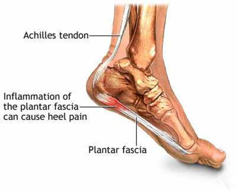
Plantar fascia
The plantar fascia is the thick connective tissue which supports the arch on the bottom of the foot. It runs from the tuberosity of the calcaneus (heel bone) forward to the heads of the metatarsal bones

Location
Actions
Ask a Question
If you have any queries relating to foot health you can ask us a question by completing the form below.
